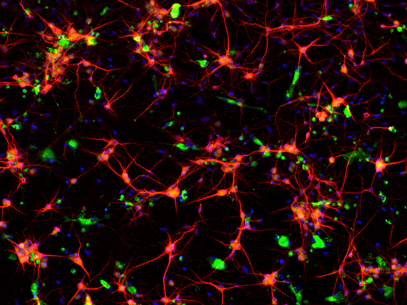
High Content Imaging Platform
Subcellular microscopic analysis to deeply detail the phenotypes (e.g., damage to neuronal morphology, subcellular organelle dysfunctions, and cytopathies) as markers of late neurodegeneration in vivo represent an important morpho-functional read-out of a CNS in vitro assay.
Bioneer uses High Content Imaging (HCI) to obtain subcellular resolution read-outs for our CNS assays.
HCI is a powerful tool in the field of drug screening, particularly for neurodegenerative diseases such as Alzheimer’s disease and Parkinson’s disease. Utilising human induced pluripotent stem cell (iPSC)-derived neurons, including dopaminergic neurons, cortical neurons, and sensory neurons, HCI facilitates comprehensive phenotypic analysis essential for advancing drug discovery.
Bioneer’s collection of iPSCs containing many disease-relevant mutations, combined with our expertise in cell differentiation for various neuronal subtypes, microglia and astrocytes provide physiologically relevant disease models for drug development assays.
Using our advanced data processing workflow and Bioneer’s proprietary image analysis software we can investigate important cellular parameters, such as:
- Neurite outgrowth
- Neurite fragmentation
- Lysosomal trafficking
- Phagocytosis
- Viability
These are key parameters to understanding neurodegenerative pathology and are believed to be key drivers associated with Alzheimer’s, Parkinson’s and other neurodegenerative diseases.
Our HCI platform, integrating human iPSC-derived neurons and advanced imaging and analysis tools, provides a robust platform for neurodegenerative disease research. As a comprehensive platform, generating valuable phenotypic data at the cellular and sub-cellular level our team can assist in unravelling complex biological process or drug candidate effects for your research project.

Co-culture (NGN2 Neurons + Microglia)
For further information
please contact:
Dale Shelton, PhD,
Director of Business

